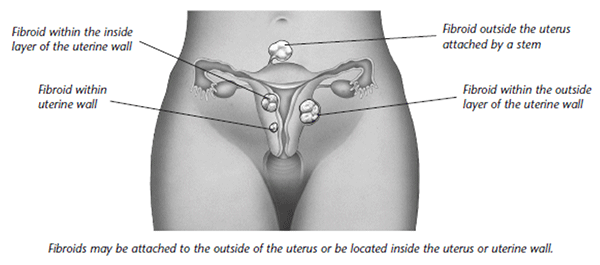
Uterine Fibroids Uterine fibroids are benign (non cancerous) growths that originate from the uterus muscle tissue. These growths are also referred to as leiomyomas or myomas. Fibroids can be found inside the uterus, within it’s wall, or on the outer surface – and also vary in size and shape. A woman could have just one fibroid or several of them in varying sizes. A fibroid can stay really small for an extended time and all of the sudden grow quickly, or get bigger gradually over a period of time. Fibroids are most commonly seen in women 30-40 years old, however they may appear at any age. Fibroids develop more frequently in African American women as compared to Caucasian women. Fibroids also seem to happen at a younger age and grow more rapidly in African American women.
Fibroids can cause these symptoms: Pain
Changes in menstruation
Pressure
Enlarged uterus and abdomen Miscarriages Infertility Fibroids can also produce no symptoms whatsoever. Fibroids are sometimes discovered during a regular scheduled pelvic exam or in the course of testing for other concerns. Fibroids which are connected to the uterus with a stem can twist which can trigger nausea, pain or fever. Fibroids that grow quickly, or the ones that start wearing down, may also produce pain. They are seldom connected with cancer. A really large fibroid might result in abdominal swelling making it difficult to perform a comprehensive pelvic exam. The initial indications of fibroids might be discovered during a routine pelvic exam. Numerous tests may reveal more details about fibroids:
|
||